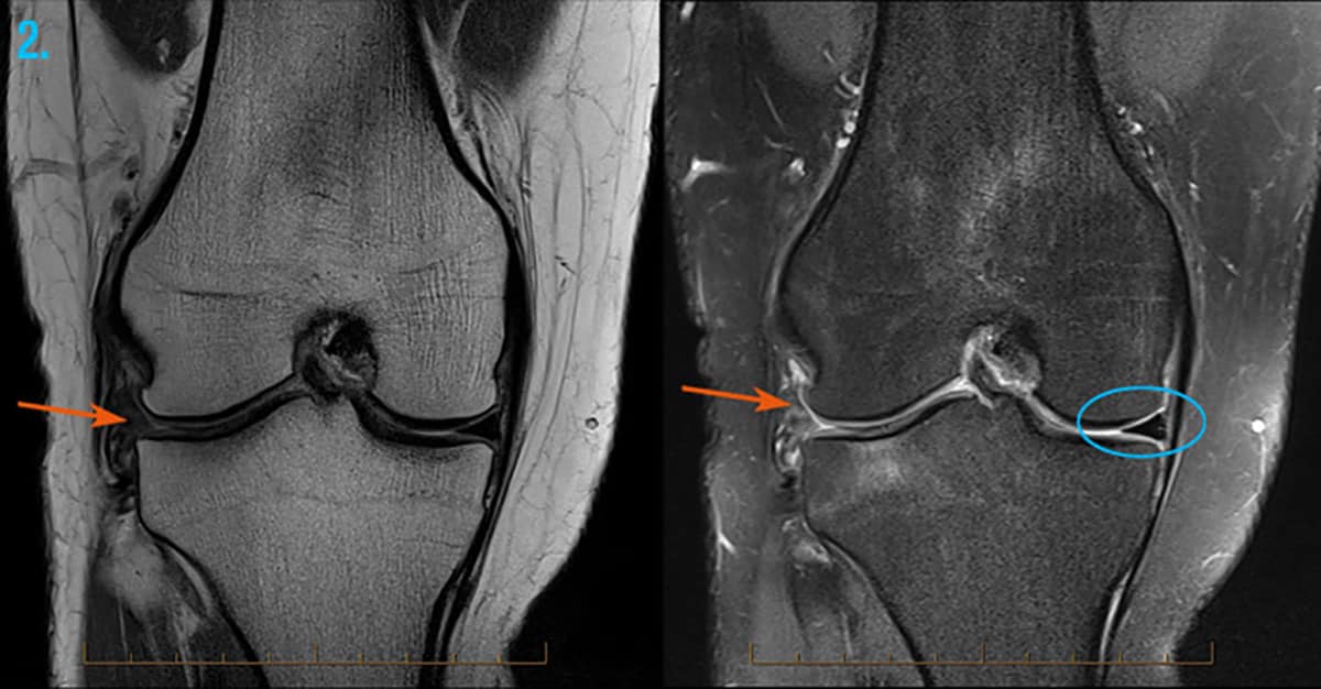
Double PCL sign - appears on sagittal MRI images of the knee when a bucket-handle tear of a meniscus most commonly the medial meniscus flips into the intercondylar recess and comes to lie. It works by creating a magnetic field that causes the water molecules in tissue bones and organs to orient themselves in different ways.
:background_color(FFFFFF):format(jpeg)/images/library/13476/mri-axial-knee-femoral-condyles-3_english.jpg) How To Read The Normal Knee Mri Kenhub
How To Read The Normal Knee Mri Kenhub
An MRI machine uses a magnetic field and radio waves to create pictures of internal organs and bone structures.

Knee mri images. They were created by volume rendering from a CT-scan of the knee. Trouvez les Knee Mri images et les photos dactualités parfaites sur Getty Images. Coronal and axial proton density images with fat saturation.
In this modality fat and hyaline cartilage show as white bones as white to gray muscles as gray and tendons and ligaments as black. Quick patient focused tutorial on how to read a knee MRI by Dr. These illustrations review the basics of the anatomy of the knee and make it easier to place them on an MRI by using the osseous cross-references.
Although there is no definitive consensus on the type of images in daily practice spin echo images with a short TE and particularly PD images are the most widely used. Magnetic resonance imaging of knee injuries in children. Magnetic resonance imaging MRI is a technology often used to investigate the sources of knee problems.
This MR image of the knee shows femur patella tibia and menisci. These orientations are then translated into images we can use for diagnosis. While a detailed explanation of MRI protocols and MR physics is beyond the scope of this text fast spin echo FSE MRI is most commonly utilized for MRI of the knee.
Chris Centenoknee mri howto. Separate knee MRI image with anatomical topical structures of that specific area muscles ligaments tissues and cartilage area of knee joint. It can be performed on any part of your body.
26287290 4Crues JV 3rd Mink J Levy TL Stoller DW. Ultimately the image produced by the MRI is a thin slice through the knee in one of these three planes. We added 3D illustrations of the bones of the knee femur tibia fibula peroneal and patella kneecap which we then labeled.
Osteoarthritis hip and knee MRI. As an example shown here is an overview of the knee on an axial PD image on a slice through the femoral condyles. If the hospital has an open MRI machine it will often be used for a knee MRI for patient comfort.
Choisissez parmi des contenus premium Knee Mri de la plus haute qualité. As a general rule MRI examination of the knee includes T1-weighted or PD proton density-weighted sequences in the sagittal plane without fat saturation followed by PD images with fat saturation in the 3 spatial planes. This MR image of the knee shows femur patella tibia anterior and posterior cruciate ligament.
Images are obtained at a 3-mm slice thickness with a Carty HM Brady O. If the MRI machine is closed the MRI table will slide inside the machine before it starts. Specifically in the coronal and sagittal planes T1 T2 and intermediate-weighted proton density FSE.
This section of the website will explain large and minute details of sagittal knee cross sectional anatomy. 3D osseous model of the knee. Medical images from an MRI allow medical professionals to distinguish body tissues including the meniscus shock absorbers in the knee cartilage tendons and ligaments.
Conceptual photo about Knee MRI Magnetic Resonance Image with written text. Sagittal Knee MRI Images T1 Weighted This T1 weighted MR image of the knee shows femur tibia and meniscal tear of the posterior horn of the medial meniscus. Medical technical assistant preparing scan of knee with MRI.
Trouvez les Mri Knee images et les photos dactualités parfaites sur Getty Images. Once the patient is ready she or he is helped onto an MRI table and pillows are used to brace the knee and hold it in place. Three conventional MRI planes that are utilized to evaluate the knee include sagittal oblique coronal and transaxial planes.
Magnetic resonance imaging MRI is the modality of choice for evaluating the soft tissue structures of the knee and indeed the knee is the most commonly imaged joint using MRI. An MRI test uses magnets and radio waves to capture images inside your body without making a surgical incision. Choisissez parmi des contenus premium Mri Knee de la plus haute qualité.
Magnetic resonance imaging MRI is a radiologic procedure that uses a magnetic field and radio waves to develop detailed image cross-sections of the body including the knee 1.
 5 Minute Knee Mri It S Possible
 Normal Knee Mri 10 Year Old Male Radiology Case Radiopaedia Org
 Mri Scan Knee Melbourne Melbourne Radiology Clinic
 Magnetic Resonance Imaging Mri Defined Cartilage Degeneration And Joint Pain Are Associated With Poor Physical Function In Knee Osteoarthritis The Oulu Knee Osteoarthritis Study Osteoarthritis And Cartilage
 Normal Knee Mri Radiology Case Radiopaedia Org
 Diagnostic Performance Of 3d Tse Mri Versus 2d Tse Mri Of The Knee At 1 5 T With Prompt Arthroscopic Correlation In The Detection Of Meniscal And Cruciate Ligament Tears
 Why Might I Need A Knee Mri Scan
 Patients Ask Is My Mri Accurate Caring Medical Florida
 Normal Knee Mri Scan Stock Image C026 1158 Science Photo Library
 Normal Mr Imaging Anatomy Of The Knee Magnetic Resonance Imaging Clinics
 Pin On Anatomy Imaging
 Knee With Multiple Abnormalities On Mri Indicating Early Stage Download Scientific Diagram
 Knee Mri Affordablemri Com
How To Read Knee Mri Of Radial Meniscus Tear Sports Medicine Knee Specialist Twin Cities Mn Youtube


Comments
Post a Comment